Bandpass filters are essential optical components in multiple applications, including remote climate monitoring, biomedical, planetary exploration, commercial imaging and product quality inspection, autonomous car navigation, and reconnaissance and surveillance. Other applications utilizing filters include archeology, geology and planetary, and asteroid surface topography.
In this issue, we will concentrate on filters for medical research and diagnostics. We will briefly review some of the roles that precision bandpass filters play to parse, measure and monitor essential properties to aid in the detection, diagnosis, tracking, and conquering of the coronavirus.
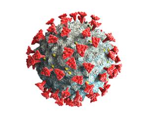
Biochemical techniques based on the analysis of blood serum are frequently used for detection and progression monitoring of the coronavirus 2 (SARS-CoV-2), which is the etiological agent of the COVID-19 pandemic. Optical techniques that rely on spectral signals isolated by precision filters find immediate application to the COVID-19 pandemic. Figure 1, based on SEM images, is a model of the 50 – 100 nm body and shows the 20 nm tall spikes (of the “crown”) that attach to and destroy respiratory cells.
Optical techniques vary in their sensitivities and detection rates and are complementary in the clinical setting. Fluorescence microscopy (FM), chemiluminescent antiviral immune response (CLIA), flow cytometry (FC), and surface plasmon resonance (SPR) are several optical techniques used for antibody immunoassay and detection of DNA and RNA strands. The detection basis is the presence of fluorochromes added or generated in the test specimen.
Fluorochromes are stains designed to attach themselves to cellular material. When illuminated, the DNA/RNA and receptors/structures emit a quantifiable quantum yield. FM and its sister technology, FC, both require separation and high isolation of excitation and emission wavelengths to illuminate, detect, and reconstruct the cells or material(s) on the slide with precision and accuracy. FM images the spatial distribution of reaction sites, whereas the multi-parameter FC technique can assay reacted cell population and characteristic fluorescent emissions.